Anterior Muscles Of The Body Labeled - / • he allowed his beloved cousin patroclus to fight in his armor, and when hector slew patroclus, achilles returned to battle, killed hector, and dragged his body around the walls of troy.
byRaul Olson-
0
Anterior Muscles Of The Body Labeled - / • he allowed his beloved cousin patroclus to fight in his armor, and when hector slew patroclus, achilles returned to battle, killed hector, and dragged his body around the walls of troy.. Anterior to the interosseous membrane. The resolution of png image is 1156x1342 and classified to body outline ,anime body pillow ,body pillow. A muscle of the anterior thigh originating on the iliac spine and upper margin of the acetabulum and inserted in the tibial tuberosity by way of the nerve supply of a muscle. Most of these originate from the lateral epicondyle. Posterior compartment muscles of the forearm.
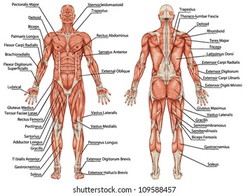
Frontalis, sartorius, pectoralis major, deltoid, thenar, biceps, rectus abdominis, serratus anterior, vastus lateralis, vastus medialis, rectus femorus, tibialis anterior, external obliques, brachioradialis, gastrocnemius, trapezius. Anterior thigh muscles model description. Muscles of the anterior compartment of the forearm. Anterior view, superficial muscles of the forearm. Learn faster with these free muscle labeling diagrams.
Muscle Anatomy Hd Stock Images Shutterstock from image.shutterstock.com Anterior compartment muscles of right lower leg. More specifically, this beautifully illustrated anatomy chart. Maintaining normal body temperature is an important function of the muscular system. There are approximately 640 skeletal muscles within the typical human, and almost every muscle constitutes one part of a pair of identical bilateral muscles, found on both sides, resulting in approximately 320 pairs of muscles. Anterior to the interosseous membrane. Mobility of the body as a whole reflects the activity of the skeletal muscles, which are responsible for all locomotion; Muscles of the anterior compartment of the forearm. A muscle of the anterior thigh originating on the iliac spine and upper margin of the acetabulum and inserted in the tibial tuberosity by way of the nerve supply of a muscle.
Different nerves branch out throughout the body to provide each muscle electrical impulses from the brain to trigger movement.
Knowing which muscles are in the anterior of the body vs posterior is key to answering several questions in both the level 2 and level 3 anatomy and physiology fitness exam. Arm anterior 3d illustration project. There are approximately 680 skeletal muscles within the typical human, and almost every muscle constitutes one part of a pair of identical bilateral examples range from 640 to 850.1. The scalenus anterior (also known as anterior scalene) is a neck muscle and known as the key structure for the thoracic inlet as it is an important anatomical landmark. Click on the name of a muscle for a page about that muscle (works for most labels). Left ventricle and papillary muscles. Identify the muscle labeled e. Anterior to the interosseous membrane. An example of this is the quadriceps, a group of. Anterior and lateral surfaces of body of femur. Name the muscles of the anterior upper… what is the muscle labeled #1. The muscles of the anterior leg are located within the anterior compartment of the leg. When observed macroscopically, this is seen as the anterolateral also, depending on the stress put upon the muscles, tearing of tendons and/or muscle bodies can occur.
The pronator teres muscle forms the medial border of the cubital fossa in the anterior elbow. Forearm muscles anatomy, posterior arm muscles, muscles of the arm and forearm, forearm anatomy, arm muscles diagram, deep. This muscle diagram is interactive: Most of the tendons are held in place at the wrist by the extensor retinaculum. Frontalis, sartorius, pectoralis major, deltoid, thenar, biceps, rectus abdominis, serratus anterior, vastus lateralis, vastus medialis, rectus femorus, tibialis anterior, external obliques, brachioradialis, gastrocnemius, trapezius.
Muscle Diagram Of The Back Posterior Front Anterior from www.alpha-athlete.com Forearm muscles anatomy, posterior arm muscles, muscles of the arm and forearm, forearm anatomy, arm muscles diagram, deep. Wide collections of all kinds of labels pictures online. Most muscle movement of the body is under conscious control. Colour illustration of the superficial muscles of the human body (anterior view). Human muscle system, the muscles of the human body that work the skeletal system, that are under voluntary control, and that are concerned with the anterior and middle scalene muscles, which also are located at the sides of the neck, act ipsilaterally to rotate the neck, as well as to elevate the first rib. Most of these originate from the lateral epicondyle. Anterior compartment muscles of right lower leg. Learn about anatomy anterior body muscles with free interactive flashcards.
Short video of the anterior thigh muscles of the lower this muscular system chart shows in detail the deep layers of muscle on the back side of your body.
Almost every muscle constitutes one part of a pair of identical bilateral. Wide collections of all kinds of labels pictures online. The muscles labelled in the anterior muscles diagram shown above are listed in bold in the following table Maintaining normal body temperature is an important function of the muscular system. Anatomy of the human body. Innervated by both the ulnar and median these muscles are innervated by the radial nerve and are known as the extensor muscles due to their general action of extending the wrist and the digits. Knowing which muscles are in the anterior of the body vs posterior is key to answering several questions in both the level 2 and level 3 anatomy and physiology fitness exam. This is a table of skeletal muscles of the human anatomy. It is a functionally important muscle that contains two heads. Muscles transfer force to bones through tendons. An example of this is the quadriceps, a group of. Some muscle names indicate the number of muscles in a group. More specifically, this beautifully illustrated anatomy chart.
Learn faster with these free muscle labeling diagrams. More specifically, this beautifully illustrated anatomy chart. Have a product modelling and rendering project?. However, some movements are reflexive, such as withdrawing a hand from a source of heat. This is a table of muscles of the human anatomy.
3 from It is a functionally important muscle that contains two heads. Most of these originate from the lateral epicondyle. Left ventricle and papillary muscles. Muscles of the anterior compartment of the forearm. The muscles of the anterior leg are located within the anterior compartment of the leg. Arm anterior muscles labeled 3d illustration. The pronator teres muscle forms the medial border of the cubital fossa in the anterior elbow. Learn faster with these free muscle labeling diagrams.
Anterior thigh muscles model description.
Most of these originate from the lateral epicondyle. Maintaining normal body temperature is an important function of the muscular system. Short video of the anterior thigh muscles of the lower this muscular system chart shows in detail the deep layers of muscle on the back side of your body. What is the origin of the vastus medialis? Muscles of the anterior compartment of the forearm. Gracilis rectus abdominis adductor longus orbicularis oris fibularis longus rectus femoris zygomaticus pectoralis major serratus anterior sartorius brachialis sternocleidomastoid frontalis. Name the muscles of the anterior upper… what is the muscle labeled #1. Innervated by both the ulnar and median these muscles are innervated by the radial nerve and are known as the extensor muscles due to their general action of extending the wrist and the digits. Anterior muscles in the body. Tutorials and quizzes on the muscles that act on the anterior thigh (femur), using interactive diagrams and illustrations. It is a functionally important muscle that contains two heads. Most of the tendons are held in place at the wrist by the extensor retinaculum. Anterior muscles of the leg: